Liver is an organ which is very well viewable by sonography. We examine it by patient lying on his back or on his left hip (better image of liver hilum). Liver is examined by fan-shape movement by probe in both sagittal and transverse plain. Liver is much more visible during inspiration.
Liver hilum (portal vein, common bile duct, hepatic artery) is displayed in right subcostal space. The probe is rotated from transverse plain for approximately 45° clockwise.
Important numbers:
Liver width: under 14cm (We measure after expiration in right sagittal medioclavicular line)
Portal vein width: under 13-15mm (We measure approximately 1.5cm before its intrahepatal bifurcation, sometimes we measure near place where portal vein enters liver parenchyma.)
Common bile duct width: under 6mm (or under 9mm after cholecystectomy)
Hepatic veins width: under 6mm for peripheral hepatic veins, under 10mm liver veins before entering the inferior vena cava.

Liver width is measured in right middle medioclavicular
line. This cut also often displays right kidney.
Liver vessels - Liver veins – Anechogenic x Portal vein branches – They have 2 hyperechogenic bright "rails" in their wall (intrahepatal bile ducts)
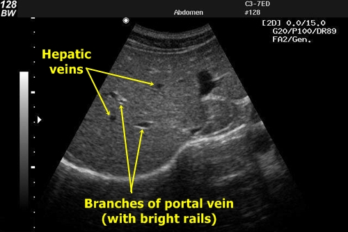
Differentiation of liver veins from portal branches
Liver hilum – It is the entrance of hepatic artery, portal vein and departure of common bile duct from liver. In this area we can measure the width of portal vein and most importantly the width of the bile duct. It is common that bile duct and hepatic artery are located above the portal vein. We can differ them by using Doppler sonography. Hepatic artery shows blood flow, bile duct does not.
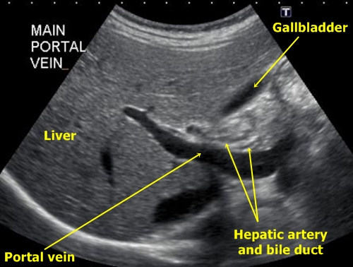
In liver hilum we see portal vein entering the liver. Two slim lines above portal vein
is hepatic artery and bile duct. In this static picture without the possibility to check
the blood flow it is practically impossible to differentiate them.
Portal hypertension – Portal vein is broader than 15mm, we also find splenomegaly and often ascites (anechogenic black fluid in abdominal cavity).
Liver congestion by right-sided heart failure – We find dialted peripheral hepatic veins over 6mm. We can sometimes find fluid in pleural cavity above the diaphragm.
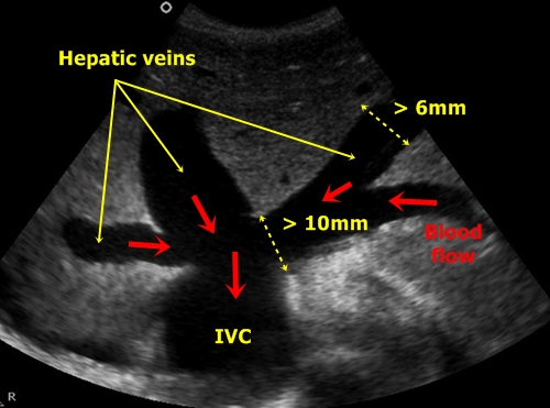
Dilated hepatic veins – Peripheral veins are broader than 6mm, veins
near influx into inferior vena cava are wider than 10mm.
Liver steatosis – The liver echogenity is higher than normal. Liver parenchyma becomes brighter than right kidney parenchyma. In sagittal projection we can easily display both organs and make a comparison.
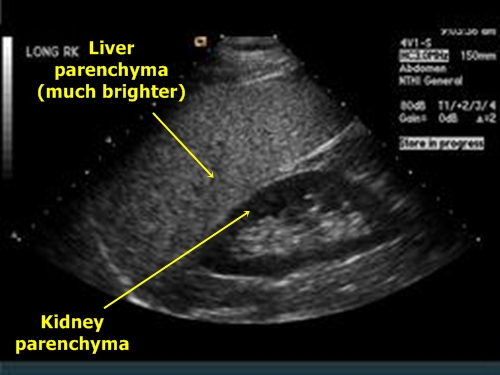
Liver steatosis – There is a clear difference between
the echogenity of liver and right kidney parenchyma
Liver cysts – It is a very common finding. They are anechogenic black round regular objects inside the liver parenchyma. Often we can see a conic bright shadow behind them (acoustic amplification).
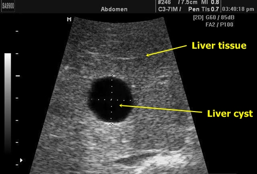
Liver cyst – It is a regular anechogenic round
structure with a conic bright shadow behind it.
Liver hemangioma – They are displayed as distinctly bordered hyperechogenic areas.
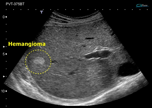
Liver hemangioma - Well bordered, hyperechogenic
Liver cirrhosis – By liver cirrhosis we find irregular rough liver margins (macronodular or micronodular cirrhosis). Liver veins connect in open angles. Parenchymal echogenity is higher and the tissue seems to be granular. In advanced stages of the cirrhosis liver size dwindles and we find marks of portal hypertension (ascites, portal vein dilation, splenomegaly).
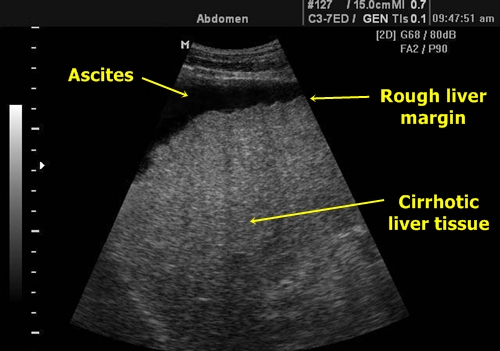
Liver cirrhosis – We see bright parenchyma of the liver,
rough liver margins and ascites in the surroundings.
Common bile duct dilation - It is a very imporntant finding and means dilation of bile duct over 6mm (or over 9mm by patients after cholecystectomy). The most common causes are gallstones and local tumours. The ultrasound, however, does not always find the cause.

Bile duct dilation – It is a pity I was not able to find such a great picture in better quality.
We see liver hilum with portal vein and dilated bile duct. In this case we also see the reason
of the dilation – bright gallstone with dorsal acoustic shadow obstructing the bile duct.
Liver tumor – It has different echogenity from surrounding parenchyma. It often oppresses neighbouring liver tissue.
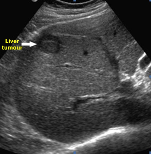
Primary tumor – This object could be also a metastasis. However,
the patient was later diagnosed with a hepatocellular liver carcinoma.
Liver metastases – They are bearings of different echogenity than liver tissue. They have typically a hypoechogenic margin (halo).
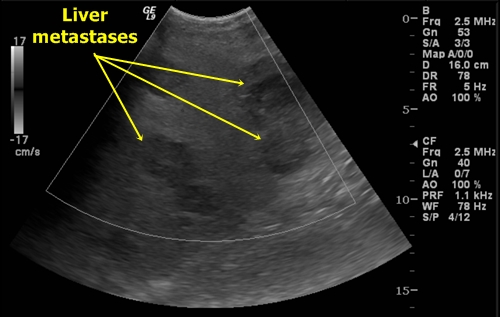
Liver metastases I – Few bigger metastases with hypoechogenic margins
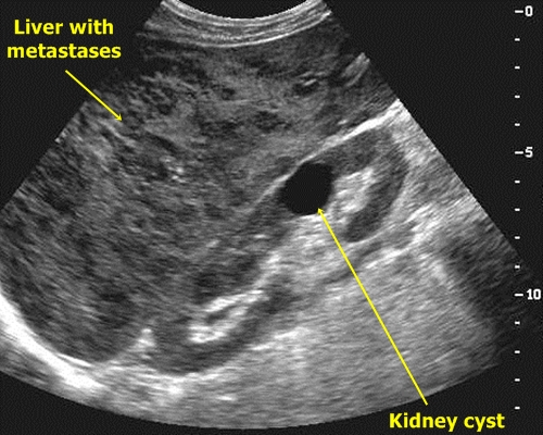
Liver metastases II – Liver parenchyma is full of small metastases. Furthermore
there is a renal cyst, which is a casual (and in this case probably insignificant) finding.


