
The inferior vena cava (IVC) can be examined in transversal and sagittal direction – the same way as the abdominal aorta. It can be well displayed in the epigastrial area where we find IVC next to the aorta in front of vertebral body. Except that we often visualize the IVC cavity near the upper part of the liver where it is entered by three main hepatic veins.
Dimensions:
Width - under 20 mm (for athletes under 25 mm) + a forced inspiration causes IVC to collapse and shrinks its diameter to about a third of its original value.
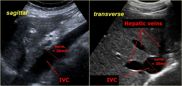
On the left we see the IVC in a longitudinal projection. The image on the right
is a cross-section of the inferior vena cava at a close contact with the liver tissue,
where IVC receives all three hepatic veins.
Right heart failure - Among other signs (see ultrasound of liver) we can find dilatation of the vena cava to diameter bigger than 20 mm (or over 25 mm by athletes). The forced inspiration causes only minor or no collapse of IVC at all.