
This is just an auxiliary text dedicated to liver functional anatomy. Anatomic orientation in liver parenchyma has a big importance for sonography. Liver segments are used for specific localisation of various pathologies in liver and for surgical liver resections. Liver is divided into eight segments I-VIII.
The segments are divided by hepatic veins and branches of portal vein. Segment I is anatomically lobus caudatus, which is located on the backside of the liver to the left from IVC.
Right Hepatic Vein - It "divides" the right lobe into anteromedial part (Segments V, VIII) and posterolateral part (VI, VII).
Middle Hepatic Vein - It is border between Segment IV (part of left lobe) and Segments VI, VII (part of right lobe)
Left Hepatic Vein - It divides the left lobe into segment IV on one side and Segments II, III on the other side.
Left Branch of Portal Vein - It divides the upper part of the left lobe (Segment II) from the lower part (Segment III).
Right Branch of Portal Vein - It divides 2 upper segments (VII, VIII) from 2 lower segments (V, VI).
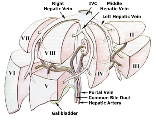
Here we can see a very illustrative picture of hepatic segments and main veins as mentioned above. Segment I is not seen, it would be located behind the IVth Segment anatomically on the left from the IVC (from our point of view on the right) (Source)
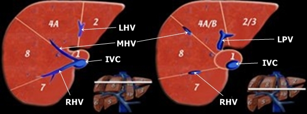
Transverse cut through the upper and middle part of the liver. RHV - Right Hepatic Vein,
MHV - Middle Hepatic Vein, LHV - Left Hepatic Vein, IVC - Inferior Vena Cava, RPV -
Right Portal Vein (branch), LPV - Left Portal Vein (branch). Liver segments are marked
by Arabic numbers. The slashes mean borders between segments. (Source)
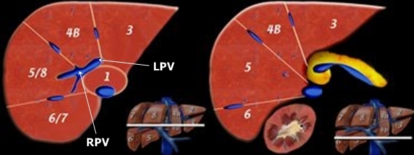
Analogy of previous picture. Transverse cuts in lower middle
and lower part of the liver tissue. (Source)
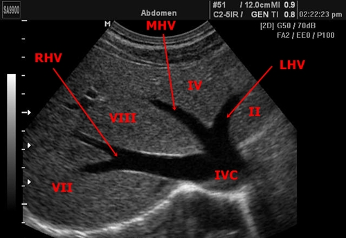
In this picture we can see marked segments in praxis. It is a slightly narrow
transverse cut in the upper part of the liver where hepatic veins flow into IVC.
Visible segments are marked by Roman numbers.