We examine kidneys in position on patients back or on the hip (right kidney on the left hip and vice versa). Both kidneys are better displayed during inspiration.
In a kidney we see hypoechogenic parenchyma with pyramids and hyperechogenic central part. We examine in both sagittal and transverse plain. Transverse cut enables us to see kidney hilum with renal artery and vein.
Proportions:
Kidney length: 10-12cm
Kidney width: 4-6cm
Parenchyma width : 13-25mm
We display right kidney in sagittal plain often together with neighbouring liver tissue. This is very important for evaluation of liver echogenity. Healthy liver tissue has the same echogenity as kidney parenchyma. Higher echogenity (which means brighter appearance) of the liver tissue speaks for steatosis.
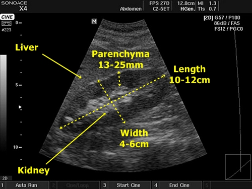
Longitudinal cut through the right kidney with marked proportions. Neighbouring liver
tissue is brighter than renal parenchyma and therefore probably steatotic.
Parenchymal bridge – This is clinically not a very important finding. It appears as a dark hypoechogenic line leading through the bright centre of the kidney.
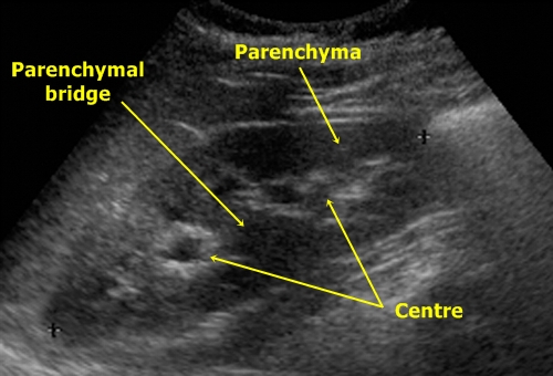
Parenchymal bridge
Dromedary hump – It is a thickened mass of renal parenchyma in left kidney near the lower part of the spleen. It is a normal finding by 10% examined patients. It could be sometimes mistaken for a tumour.
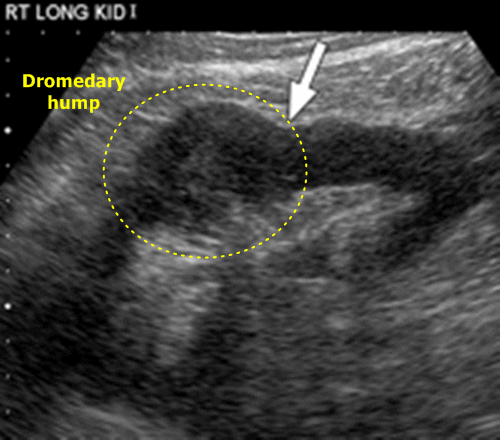
Dromedary hump - Parenchyma of the left kidney is broader in its lateral part.
Renal cysts – They are regular round anechogenic bearings. Very often we find parenchymal and parapelvic cysts. Parapelvic are located in the central part of the kidney and could be easily confused with dilatation of the pelvic tract in hydronephrosis.
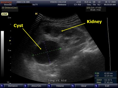
Renal cyst - It is the big round object near the kidney pole.
Pyelonephritis – Kidney is enlarged, it has higher echogenity of its parenchyma (these are non-specific signs of kidney damage which also appear by glomerulonephritis, amyloidosis, etc.).
Degenerative processes – The parenchyma becomes thinner. These processes are often connected with angionephrosclerosis. The sclerosis is displayed as short hyperechogenic lines in renal parenchyma above the pyramids.
Hydronephrosis – We find dilatation of renal pelvis and possible caliceal distension as well. It seems as large hypoechogenic objects in central part of the kidney. From dilated veins they can be distinguished by duplex sonography which would show blood flow in the veins. Differentiation from parapelvic cysts could be sometimes very difficult.
Hydronephrosis has 3 stages according to sonography:
1. Pelvic dilatation
2. Pelvic dilatation + caliceal enlargement (+ possible little parenchymal reduction)
3. Pelvic dilatation + caliceal enlargement + parenchymal atrophy
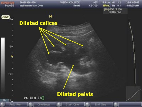
Hydronephrosis- In this picture dilatation of the pelvis and calices is visible.
However, renal parenchyma does not seem to be too atrophic.
Nephrocalcinosis – Imaging of hyperechogenic concrements is often quite difficult (sometimes nearly impossible in hyperechogenic renal centre). It is important to find hypoechogenic shadow which they create.
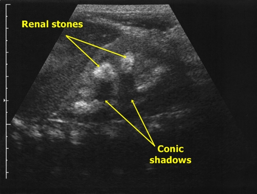
Nephrocalcinosis - We see two concrements (marked by red circles), which cannot be well distinguished from the hyperechogenic centre of the kidney. However, they form visible cone shadows.
Kidney infarctions – Kidney ischaemia appears as a triangular hypogenic lesion in parenchyma
with its basis near the renal capsule.
Kidney tumors – These are irregular anisoechogenic objects growing from the parenchyma. They can have a cystic element.
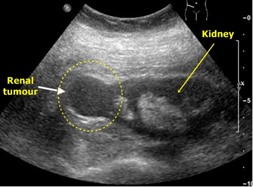
Kidney tumor -In this case quite regular mass with echogenity similar to kidney
parenchyma grows from the upper kidney pole and causes its local enlargement.
(In this case it would be easy to mistake it for a renal cyst)
Angiomyolipoma – This is a benign tumor which can be best distinguished in its first stages of growth when it looks like small hyperechogenic bearing. As it grows it becomes less echogenic and therefore more similar to malignant tumours.
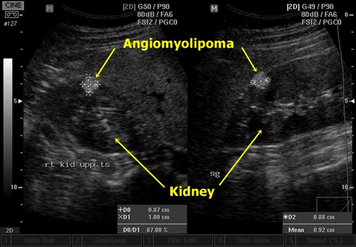
Angiomyolipoma - In two US pictures we can see
a round bright object in renal parenchyma.
