The bladder is a relatively well-viewable organ. It is essential that the patient does not pass urine before the examination. Filled bladder is visible with good assessment of its wall and we can also better see the organs located deep behind the bladder (uterus + vagina in females). Patients with urinary catheters should be plugged about two hours before the examination so the bladder will be full.
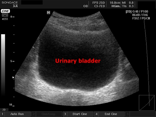
Filled bladder in the classical transverse section
Longitudinal section of the urinary bladder by a woman – behind the bladder there is vagina and uterus reaching above the bladder. More filled the bladder is the better picture we get.
Determining residue: We have to measure the urinary bladder in three diameters – the diameter in transversal plain, craniocaudal diameter in the sagittal cut and anteroposterior diameter in any of those plains. If we multiply these values (in centimeters) and then divide by 2, we get the volume of fluid in the bladder in milliliters. Physiological residue after urination is less than 100 ml.
Wall thickening – Diffuse thickened wall is usual by cystitis. Local thickening is more typical for tumors of the urinary bladder. In that case we ideally find cauliflower-like formations emerging from the wall into the cavity of the urinary bladder.
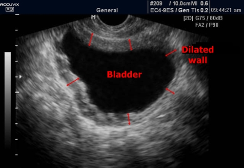
Chronic cystitis - Diffuse wall thickening (red arrows)

Bladder tumor - A suspicious cauliflower-like structure with local wall thickening
Urinary stones – They may appear as hyperechogenic formations which project classical ultrasonic shadow.
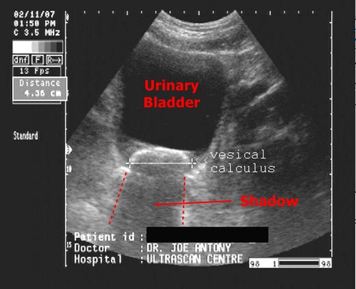
Calculus - Vesical calculus is located by the wall of the
urinary bladder. Below you can see the cone shadow.
Urinary catheter – Inside the bladder there should be an inflated catheter balloon visible as a spherical object. If the patient’s catheter has not been plugged some time before the examination the urinary bladder would be logically empty, collapsed and very badly viewable.
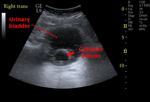
Urinary catheter – A regular spherical object in the urinary bladder marked by red arrow
Note: In the sagittal view at full bladder we can see the so-called Douglas pouch behind uterus and vagina. (excavatio rectouterina). By males we can identify similar pouch called excavatio rectovesicalis (between urinary bladder and rectum). In cases of suspected abdominal trauma with possible internal bleeding we look for anechogenic fluid (blood) in these locations.
